
Targetoid Hemosiderotic Hemangioma (Hobnail Hemangioma)
Overview
Targetoid Hemosiderotic Hemangioma, also known as hobnail hemangioma, is a benign vascular lesion with a characteristic targetoid appearance. Usually a central violacious papule or plaque with a surrounding ecchymotic (bruised) targetoid ring. The bruised ring may come and go but the central portion tends to persist between episodes. It can mimic a variety of other conditions raning from melanoma, Kaposi’s sarcoma, angiokeratoma, bite reaction or even angiosarcoma if growing and changing.
Histology
The principal feature are hobnail endothelial cells and positive vascular markers. HHV-8 stain can reliably distinguish from Kaposi Sarcoma.
Treatment
Surgical excision is diagnostic and therapuetic. The condition is benign so it does not require aggressive treatment.
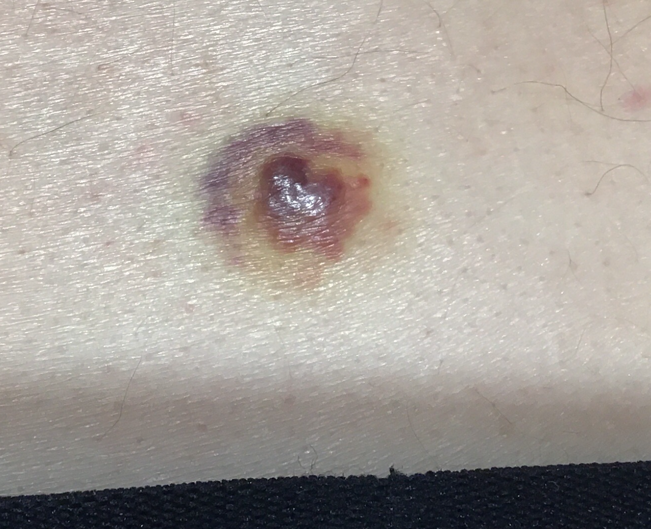
Targetoid hemosiderotic hemangioma (hobnail hemangioma) on the back.